Antibody Production In Different Host Systems: From Rabbit to Mouse
Sep 19th 2023

To identify molecules of interest in research and diagnostic settings, scientists can use antibodies produced by animal immune systems to bind precisely to antigens as a starting point for manufacturing probes. No other technology currently available enables scientists to develop and produce tools for molecular recognition that are as highly specialized as antibodies.
Most medical or cell biology researchers who conduct any molecular testing employ antibody technology in some capacity. Different researchers will spend more or less time on antibody synthesis and purification, using other hosts depending on their demands.
During the 1970s and 1980s, antibody production methods were created for making, purifying, and changing antibodies for antigen-specific probes. These methods have mostly stayed the same since Harlow and Lane published their classic Antibodies: A Laboratory Manual in 1988.
However, animal hosts have also risen in popularity over the past years.
This guide discusses antibody production in different hosts, especially rabbits and mice.
The Rising Popularity of Rabbit Hosts vs. Mouse Hosts
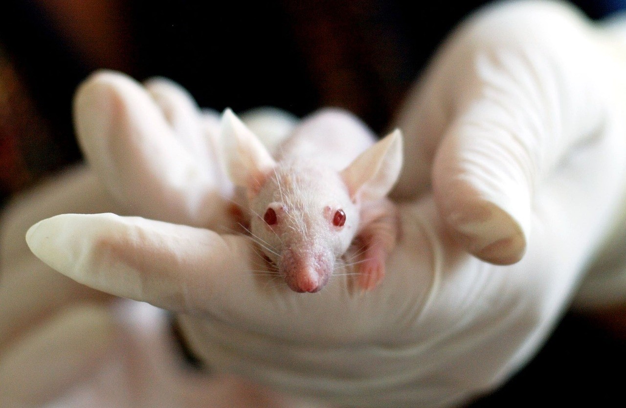
Image by Tibor Janosi Mozes from Pixabay
Since the hybridoma joined other antibody production techniques after it was first discovered in the 1970s, mice have become the primary host systems for synthesizing antibodies. However, as researchers have extended our understanding of the immune systems of various animals, other hosts have garnered interest thanks to their distinctive qualities, as opposed tomouse as a host for antibody production.
Scientists first became interested in these hosts when they found out rabbits could be viable alternatives. In contrast to rodents' immune systems, rabbits appeared to be able to identify a significantly wider variety of antigens. Surprisingly, the products of rabbit antibody production have a much higher affinity, particularly for epitopes that come from humans or epitopes identified as non-immunogenic in mice.
These qualities, coupled with rabbits' bigger size than mice's, have rabbit as a host for antibody production, particularly efficient for study and diagnosis.
Antibody Production in Rabbits and Immune Reaction
Photo by Google DeepMind on Unsplash
Like the immune reaction in humans, the production of antibodies in rabbits is caused by the intricate relationship of antigen-presenting cells (APC), T cells, and B cells, which then change into plasma cells that make antibodies.
The first immune reaction causes the rabbit's body to make the IgM isotype. This is followed by a release of IgG (up to 20 mg/ml) or IgA (up to 4 mg/ml) afterward. So far, the IgD isotype in rabbits has yet to be matched up with anything else. In contrast to other species, rabbits' IgG isotype does not show subclass differentiation.
The rabbit antibody shares structural similarities with its mammalian and human counterparts, but their components differ. For example, rabbit IgG's N terminus and the D-E loop tend to have fewer amino acid residues. In addition, these biomolecules have a peculiar interdomain disulfide bond in their structure. Researchers speculate that this link contributes significantly to making these molecules more stable and extending their shelf lives.
In addition, it was discovered that the CDR3-loop, the third complementarity-determining area of the light chain, was noticeably lengthier in rabbits compared to its length in humans and mice. The higher binding power of these bioreagents is also attributed to this property.
In addition to the structural variations, rabbits' methods for developing their antibody repertoires are notably distinct from those documented for rodents and humans. Also, the rabbit's size and the conditions in which it grows considerably impact its varied naive repertory. For instance, newborn bunnies have a weakened immune system compared to older rabbits.
Additionally, comparative antibody production in rabbits brought up in germ-free environments has been found to produce a primary antibody repertoire that is only moderately diverse.
In contrast to humans and rodents, rabbits have a substantially smaller VH repertory, although their VL repertoire is considerably more extensive and varied. Although these hosts have a smaller VH repertoire, they often make up for it with much higher rates of gene conversion and somatic hypermutation.
Production of Polyclonal Antibodies in Rabbits as Opposed to Mice
In most cases, rabbit polyclonal antibodies are chosen above their rat counterparts for use in diagnostics and in writing analytical essays. This preference can be traced back to three primary causes, the first of which is associated with the physical size of rabbits. These hosts can produce significantly more antisera because they are far more significant than the ordinary rats employed in the manufacture of antibodies.
In addition, rabbits can produce antibodies opposing antigens that typically do not cause a reaction from the immune system of rodents. Finally, rabbits are effective at producing antisera with a high titer of antibodies and greater sensitivity and affinity towards a particular target. This is backed by extensive research.
Rabbit Monoclonal Antibody Production
Using Hybrid Technology
There is still a significant need for polyclonal rabbit antibodies as reagents. But, just like traditional polyclonal antibodies made from rodents, these products vary from batch to batch. As a result, monoclonal antibodies should be utilized instead of polyclonal antibodies for applications requiring high uniformity and repeatability.
Generating rabbit monoclonal antibodies is substantially more challenging than generating their mouse equivalents. Hybridoma technology was initially used to try and produce these antibodies. However, these efforts were unsuccessful because rabbit spleen cells did not have access to a suitable fusion partner at the time.
Numerous studies were conducted on the early hybrid cell lines of mouse-rabbit heterohybridomas. However, it was discovered that they were prone to instability and were challenging to clone. A study conducted in 1995 at Loyola University in Chicago (United States) and directed by Katherine Knight was responsible for the first significant advance in the field.
The researchers used a double-transgenic rabbit that overexpressed the v-abl and c-myc oncogenes to create a compatible fusion partner for rabbit spleen cells. The transgenic rabbit got a growth that looked like a myeloma, which made it possible to separate the 240E-1 cell line.
After that, researchers fused the 240E-1 cell line with cells from rabbit spleens. However, the resulting hybridomas were genetically prone to instability and rapidly losing the genes that encoded antibodies. Weimin Zhu and Robert Pytela, scientists at the University of California, San Francisco (USA), eventually found solutions to the stability issues. These scientists chose to subclone 240E-1 multiple times and optimize the medium to stabilize it.

Image by Arturs Budkevics from Pixabay
As a direct consequence of this, the progeny of 240E-1 displayed improved fusion efficacy and robust development. The top-performing offspring clone was designated 240E-W. The union of these immortal cell lines with cells from rabbit spleen produced many stable hybridomas, which were shown to sustain steady growth without compromising their viability.
Further research using the 240E-W cell line resulted in major advances in stability and fusion performance. The expression of indigenous rabbit heavy and light chains was silenced in subsequent cell lines, 240E-W2 (US Patent 7,429,487) and 240E-W3, produced in additional research to boost the efficiency of fusion reactions and enhance the manufacture of antibodies.
Phage Display Technology
The use of phage display for choosing antibodies was first documented in the early 1990s. On the other hand, Christoph Rader and other researchers from The Scripps Research Institute in the United States did not report the use of the technology in rabbit repertoires until 2000. In this research, the scientists used the robust approach to discover and humanize binders towards the human A33 antigen from a naive scFv rabbit-derived library.
Because of the challenges in generating rabbit hybridomas, phage display technology emerged as the preeminent method for producing rabbit monoclonal antibodies. Whereas the Fab section of rabbit antibodies can also be used for phage display, the scFv rabbit/human Fab format is now the most popular choice.
Mouse Antibody Production (Monoclonal)
The primary principle behind creating monoclonal antibody generation in mice is that each antibody-producing cell only generates a single, highly specific antibody. For antibody production in different hosts, it is necessary to induce a polyclonal response caused by immunization. This is because vaccination synthesizes multiple antibodies, each with a unique sensitivity and isotype. These cells have a short lifespan.
Thus, they are crossed with a myeloma cell line that does not produce antibodies to develop a hybrid that does. Because cancer cells are enduring, researchers have developed a technique called hybridoma technology to combine cancer cells with healthy ones.
After the mouse has been immunized with the target antigen, its spleen will be harvested once it has been established that polyclonal antibodies have been successfully produced. These spleen cells have been chemically merged with a myeloma cell line. The hybrid cells are then hand-picked and given a chance to thrive.
However, because of the poor success rate of selecting cells that produce the necessary antibodies, a selective medium is used where only fused cells may develop.
This can occur since myeloma cells have lost the ability to produce an enzyme called hypoxanthine-guanine-phosphoribosyl transferase ( HGPRT), which is necessary for nucleic acid salvage synthesis. However, these cells cannot proliferate without a deficiency in HGPRT or the pyrimidine production process.
When cells are confronted with aminopterin, a folic acid derivative that blocks dihydrofolate reductase ( DHFR), they can't go through the de novo route. They can't make their nucleic acids, so they need to be fed extra nucleic acids.
Hypoxanthine, aminopterin, and thymidine are the three components in the vitamin-lacking culture medium referred to as HAT medium. This medium is optimal for the proliferation of hybridoma fusion cells. However, myeloma cells that have not fused can't continue to grow since they are missing the HGPRT gene, making them unable to duplicate their DNA.
Unfused spleen cells can only live for a brief period, which limits their growth potential. Meanwhile, hybrid cells, referred to as fusion hybrids, have the potential to develop endlessly when cultured. The myeloma companion cell possesses features that render it immortal, while the spleen cell offers HGPRT.
To help simplify the procedure of monoclonal antibody production in mice, you check the next section.
Typical Steps Involved in Monoclonal Antibody Production in Mice
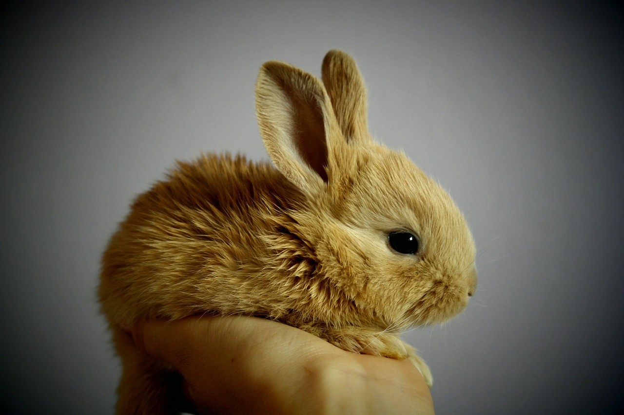
Image by Simona Robová from Pixabay
Step #1:
Mice Immunization and Spleen Cell Isolation - Mice are given an antigen to be immunized against, and then their blood is analyzed to check for the formation of antibodies. In the lab, the splenocytes that make antibodies are then used to make hybridomas.
Step #2:
The process of preparing myeloma cells - Myeloma cells are permanent cells that, when joined with cells from the spleen, can form a hybridoma that can grow indefinitely. Fusion-ready myeloma cells are created.
Step #3:
Fusion - With polyehthylene glycol (PEG) available, cell membrane fusion is induced where myeloma cells and isolated splenocytes are combined to generate hybridomas. This process takes place in the laboratory.
Step #4:
Clone detection and selection - Clones are examined and chosen based on their sensitivity to antigens and their immunoglobulin class.
Step #5:
Characterizing functions - Each high-yielding colony is verified, validated, and described (using techniques like ELISA test kits).
Step #6:
Scale up and Screen -It is recommended to increase the number of clones that can produce the desired antibodies and then gradually stop using the selection agent(s).
Step #7:
Expansion - Increase the number of clones producing the desired antibodies using various methods (such as bioreactors or huge flasks).
Wrapping Up
Similar to the case with murine antibodies, rabbit monoclonal antibodies frequently require humanization before their use in therapeutic settings. The same techniques used to humanize antibodies made from mice have also been used to humanize antibodies after successful antibody generation in rabbits.
It is already conceivable to graft all six of a rabbit's CDRs into a human IgG framework. This can be accomplished by either rational design, guided evolution, or combining the two approaches. These two host organisms for antibody production are still far from perfect.
Consequently, just a single rabbit-derived antibody ( Brolucizumab) has been authorized in humans. This is why research, diagnosis, and analysis remain the most common uses for these antibodies. Therefore, it’s vital to obtain them from a trustworthy supplier.
So, suppose you’re looking for antibody sequencing companies,protein production companies, Elisa kit manufacturers, peptide synthesis companies, recombinant antibody services, or even peptide synthesis prices. In that case, you can contact Biomatik today to speak with a specialist.